Understanding The MRI Scan- How It Works & What To Expect
Magnetic resonance imaging, or MRI, is a test that doctors use to diagnose critical conditions. The scan uses magnets & radio waves to capture internal images of the body. MRI procedure does not involve any surgical incision. The detailed images allow the doctor to see the body's soft tissues, such as muscles and organs. MRI has proven its value in detecting a broad range of severe conditions, including cancer, heart & vascular disease, and muscular and bone abnormalities.
Detailed MR images help doctors to diagnose and detect the disease. It is a noninvasive technique involving no exposure to radiation waves.
The images of the full body MRI scan show the soft-tissue structures of the body—such as the liver, stomach, heart, uterus, and many other organs without the obstruction of the bones. MR images give an accurate identification of diseases, thus making MRI an invaluable tool in early diagnosis.
Head MRI, Also Known As MRI Brain Scan
MRI brain scan or Head MRI is a painless test that creates images of the brain and brainstem using a magnetic field & strong radio waves. This scan is also known as cranial MRI.
MRI brain scans effectively detect abnormalities in small brain structures, such as the pituitary gland. A contrast agent is administered through an intravenous (IV) line to visualize specific abnormalities better.
A pelvic MRI test can assist the doctor to see the bones, organs, and blood vessels. The pelvic area between the hips holds the reproductive organs and other critical muscles. A Pelvic MRI can diagnose hip pain or investigate certain cancers.
To know more about MRI pelvis cost, please visit our clinic. Our healthcare assistants will guide you further.
Why Is MRI Done?
Physicians use an MR test to help detect or monitor treatment for many health conditions.
● MRI of the whole body evaluates all the organs in the abdomen. The liver, kidneys, spleen, bowel, pancreas, and adrenal glands, even the pelvic organs, and the reproductive organs.
● It can diagnose blood vessels and lymph nodes.
● It can detect tumors in the pelvis.
● MR images can diagnose liver diseases such as cirrhosis and abnormalities of the bile ducts & pancreas.
● It can help to assess a fetus in a pregnant woman.
A pelvic MRI scan is a valuable test to detect the following:
● Any congenital disabilities in the person
● An injury or trauma in the pelvic area
● If the person is experiencing excessive pain in the lower abdomen
● If a person is experiencing unexplained difficulties during urinating or defecating
● MR images can detect cancer in the reproductive organs, bladder, rectum, or urinary tract.
Pelvic MRI scanning for women can help to investigate:
● If a woman has an infertility problem
● If she has an irregular vaginal bleeding
● If there are lumps or masses in the pelvic area, such as uterine fibroids
Doctors may conduct a pelvic MRI in men to look for conditions like
● If there is an issue with an undescended testicle
● If there are lumps formed in the scrotum or testicles
● If the person has pain or swelling.
How Does The MRI Procedure Work?
Our body is made up of water, and water contains hydrogen atoms. MRI machines use a high-power magnet to align the hydrogen atoms within the body temporarily. The aligned particles produce faint signals, which the machine records as images. Different amounts of energy are emitted depending on the type of tissue. The scanner captures this energy and creates a picture using a computer.
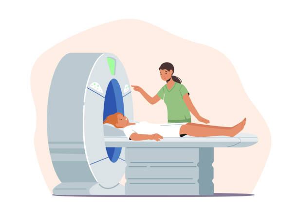
An MRI machine looks like a giant tube with a table that slowly glides you into the center. The person must lie on the table, which slides into the machine. MRI machines produce a magnetic field by passing an electric current through the coils. The coils send & receive radio waves, producing faint signals. The patient does not come into contact with the electric current.
How Is A Pelvic MRI Conducted?
The person has to lie on their back on the table that slides into the large MRI machine. A pillow or a blanket is provided to make the person comfortable. No metals are allowed in the scan room.
The radiologists will place small coils around the pelvic region to get more detailed and clear images. If the situation demands, he may put one of the coils inside the rectum to get a clearer view of the prostate or rectum. The person should lie still during the exam.
The radiologist will be in another room from where he will control the movement of the bench. The patient may communicate with him over a microphone. The radiologist may ask you to hold your breath for some seconds to take clear pictures. The person lying inside the machine does not feel any radio frequencies.
If Contrast Material Is Injected:
If the MRI exam needs contrast material, an intravenous catheter or an IV line is inserted into a vein in your arm. The contrast material helps to get detailed images. The person may feel warm or get a strange taste in her mouth when you receive the contrast injection.
MRI machines make loud whirring and thumping noises when they take multiple scan images. The patients may be provided with earplugs or headphones to decrease the noise.
MRI- A Painless Body Imaging Technique
MRI involves no incision, so it is a painless procedure. However, some patients may find lying inside the machine uncomfortable. Others may feel claustrophobic. The person may tell the radiologist about their condition.
It is normal to feel slightly warm when the contrast material is injected into your vein. If it is uncomfortable for you, inform the radiologist. The person will need to keep the same position without moving. A slight movement will distort the images.
The person is alone in the scanning room. However, you can speak with the radiologist by a two-way intercom. A squeeze ball is provided to the patients to alert the technician if the person needs any help.
What Are The Risks Involved?
The MRI test poses no risk to the patient, but you should follow specific guidelines that should be followed before and during the scan.
The strong magnetic field does not cause any harm to the person. But it may cause implanted devices to malfunction or distort the scanned images.
If you can hold your breath, the quality of the pictures will be more transparent.
The contrast material may cause little complication. However, it is scarce that any difficulty can arise. There can be problems if someone has severe kidney disease. In this case, inform the radiologist and doctor before the MRI test with contrast injection.
Will There Be An Allergic Reaction?
There is little risk of an allergic reaction. Usually, the responses are mild and resolve on their own. You should tell your doctor if you have an allergy to certain medicines.
Medical implants and other metallic objects can pose difficulty in obtaining clear pictures. Any slight movement can have the same effect. A large amount of ascites fluid in the abdomen or pelvis can also cause a hindrance and result in low-quality MR scan images.
Conclusion
MRI tests are considered the safest scanning than X-rays as they do not use ionizing radiation waves. MRI procedure creates 3D images which are very detailed and assists the doctors in making the correct treatment. Research "MRI scan near me" to find the best clinics and the related costs.


Comments