Introduction
Skin cancer, particularly melanoma, has become a growing concern worldwide. Detecting melanoma early is crucial to improving survival rates, and mole evaluation plays a key role in this detection process. Dermatologists have long relied on traditional methods for assessing moles, but Dermoscopy Mole Evaluation in Dubai, a more recent innovation, is transforming the field. This article provides a comparative study of dermoscopy and traditional mole evaluation methods, exploring their effectiveness, differences, and impact on diagnosis.
Traditional Mole Evaluation Methods
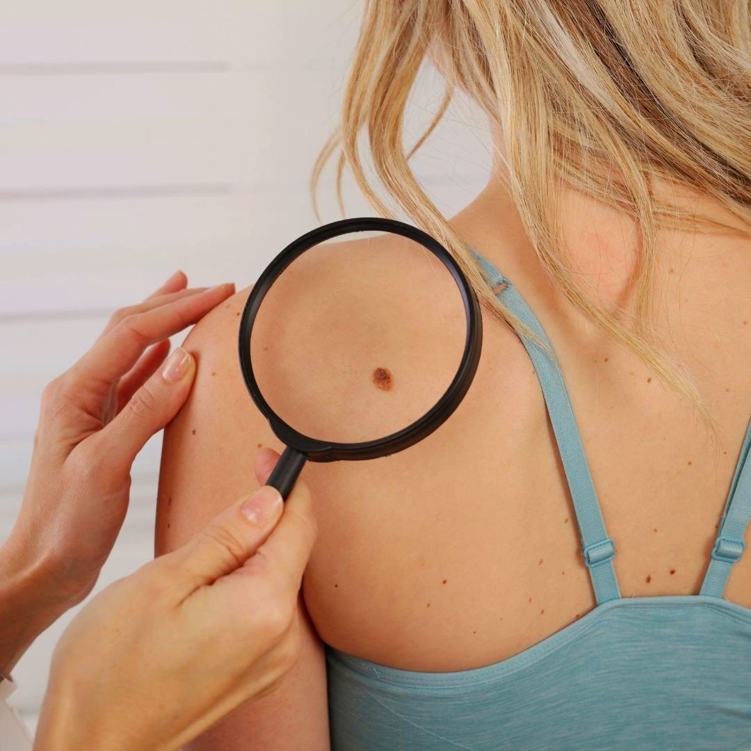
Traditional mole evaluation methods are rooted in the clinical examination of the skin. These techniques primarily rely on the naked eye and are usually based on established guidelines like the ABCDE rule. Each letter stands for a critical aspect of mole evaluation:
- A for Asymmetry: Healthy moles are symmetrical, while melanoma often appears asymmetrical.
- B for Border: Non-cancerous moles typically have smooth borders, whereas melanoma may have irregular, notched, or blurred edges.
- C for Color: Benign moles are usually uniform in color, whereas melanoma may present with multiple shades or unusual colors such as blue or black.
- D for Diameter: Moles larger than 6 mm in diameter are more likely to be malignant.
- E for Evolution: Changes in size, shape, color, or sensation over time can signal malignancy.
Doctors also rely on patient history and physical examination, noting any significant changes in moles or new growths. While this method is accessible and cost-effective, it has some limitations. For example, it is heavily dependent on the skill and experience of the clinician. This subjective nature can lead to variability in diagnosis, particularly in the early stages of melanoma, when visual characteristics may not be as evident.
Introduction to Dermoscopy
Dermoscopy, also known as dermatoscopy or epiluminescence microscopy, is a non-invasive diagnostic tool that allows dermatologists to examine the skin more closely than with the naked eye. It involves using a handheld device called a dermatoscope, which consists of a light source and a magnifying lens. By illuminating and magnifying the skin, dermoscopy provides a more detailed view of the structures beneath the surface.
The technique allows for the identification of patterns, colors, and structures within the skin that are not visible during a standard examination. These features, often referred to as dermoscopic features, include reticular patterns, pigment networks, and vascular structures. By recognizing these patterns, dermatologists can more accurately differentiate between benign and malignant lesions.
Dermoscopy has gained popularity due to its ability to improve diagnostic accuracy, particularly in the early stages of melanoma. Studies have shown that dermoscopy can significantly reduce unnecessary excisions of benign moles, minimizing patient discomfort and healthcare costs.
Comparative Analysis: Dermoscopy vs. Traditional Methods
Diagnostic Accuracy
One of the most significant advantages of dermoscopy over traditional methods is its increased diagnostic accuracy. Traditional mole evaluation heavily depends on the naked eye and often requires the clinician to make judgments based on limited surface characteristics. Dermoscopy, however, allows for a more in-depth examination by visualizing structures below the skin's surface. This deeper view enables earlier detection of malignant changes, especially in subtle cases.
Research has shown that dermoscopy improves sensitivity and specificity in detecting melanoma compared to traditional methods. The enhanced diagnostic precision reduces the number of unnecessary biopsies, which are often performed based on suspicion of malignancy. This accuracy is particularly crucial in identifying thin melanomas, which may be challenging to diagnose using traditional methods but have a better prognosis if caught early.
Training and Skill Level
While dermoscopy offers increased diagnostic accuracy, it also requires specialized training. The interpretation of dermoscopic images involves recognizing complex patterns and features that are not present in traditional evaluations. Dermatologists must undergo additional training to become proficient in using this tool effectively. This learning curve can pose a barrier for clinicians who are unfamiliar with the technique.
On the other hand, traditional mole evaluation methods require less specialized training, making them more accessible to general practitioners and healthcare providers in underserved areas. For clinicians who do not have access to a dermatoscope or the necessary training, traditional methods remain a valuable diagnostic tool.
Time and Convenience
Dermoscopy requires more time for both training and examination. Since it involves close inspection and analysis of the skin’s structures, the procedure may take longer than a simple visual assessment. While this can lead to improved diagnosis, it may be less practical in busy clinical settings with limited time.
Traditional methods are faster and more convenient for quick assessments, particularly in situations where patients present with numerous moles. This method also does not require additional equipment, making it a more straightforward and accessible option for routine check-ups.
Impact on Patient Outcomes
Dermoscopy's higher accuracy directly impacts patient outcomes by facilitating early detection of melanoma. Early-stage melanoma, when caught, can be treated more successfully, leading to a significantly higher survival rate. Dermoscopy's ability to detect melanomas that are not immediately apparent to the naked eye makes it an invaluable tool in the fight against skin cancer.
However, the increased sensitivity of dermoscopy may lead to a higher detection rate of benign lesions, potentially causing anxiety in patients. This phenomenon, often referred to as "over-diagnosis," can lead to unnecessary follow-up procedures and biopsies, which might have a psychological and financial impact on the patient.
Traditional methods, while less accurate, are generally quicker and cause less alarm in patients. However, they carry the risk of missing subtle melanomas, potentially delaying treatment and leading to worse outcomes for some individuals.
Conclusion
Both dermoscopy and traditional mole evaluation methods offer valuable tools for detecting skin cancer. Traditional methods, such as visual examination based on the ABCDE rule, remain widely used due to their simplicity and accessibility. However, these methods have limitations, especially in identifying early-stage melanomas.
Dermoscopy, with its advanced imaging capabilities, offers improved diagnostic accuracy and earlier detection of melanoma. While it requires specialized training and may be more time-consuming, its benefits for patient outcomes are significant. In conclusion, a combination of both approaches, depending on the clinical setting and available resources, can provide the best strategy for early melanoma detection.


Comments