CD BioSciences, a US-based biotechnology company focusing on the development of imaging technology, announced the availability of its new services based on Single Molecule Fluorescence Imaging technology. This breakthrough technology enables scientists to study the behavior and function of individual molecules, providing unprecedented insights into biological processes.
Traditional biological research techniques focus on studying the activity of molecules or cells. However, this approach ignores the specificity of individual molecules or subpopulations within a cluster. Indeed, the activities of individual molecules or cells at different stages of the cell cycle or in different environments are likely to differ from the overall activities of the cluster. With the advent of single molecule fluorescence imaging, researchers can gain insights into the behavior of individual biomolecules in their natural environment and their role in cellular processes.
CD Biosciences has been developing fluorescent probes and imaging technologies for many years. The company now provides customers with single molecule fluorescence microscopy to study the structure of individual molecules and their functions in physiological activities such as DNA/RNA-protein interactions, DNA replication and transcription, cell wall synthesis, and mitochondrial protein dynamics.
The new single molecule fluorescence imaging technology can be divided into two groups, one of which is the study of the activity of individual molecules in response to external forces. The mechanical properties of single biomolecules and the interactions between biomolecules are typically studied using atomic force microscopy (AFM), optical tweezers (OT), or magnetic tweezers (MT). The other is the use of fluorescence microscopy to observe the activities of single molecules in biological systems. These two types of methods provide molecular information from different perspectives and are therefore complementary.
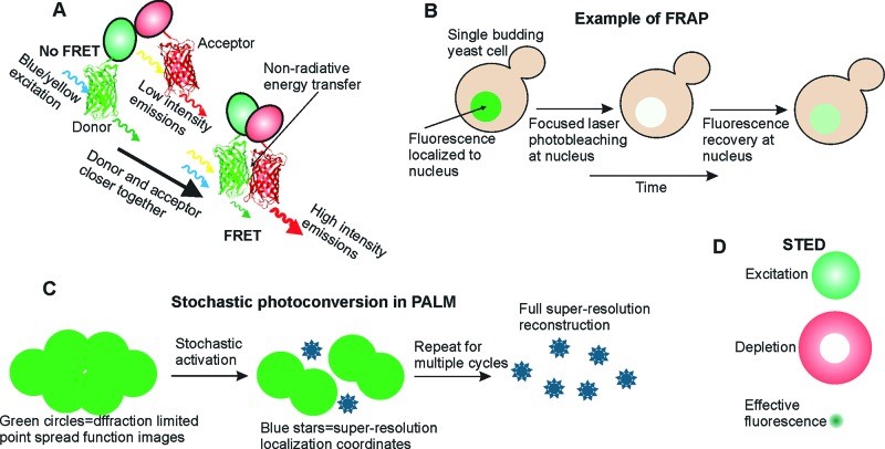
This single molecule fluorescence imaging technology is based on labeling molecules of interest with fluorescent dyes, which are then imaged and analyzed using a fluorescence microscope. Therefore, the choice of labeling method and fluorescent dye is very important and must be selected based on the requirements of the sample and the particular imaging technique. There are many methods for labeling single molecules, including antibody labeling, biotinylation, epitope labeling, small molecule probes, and bioorthogonal labeling. Common fluorescent labels include organic dyes (e.g., FITC, TRITC), fluorescent proteins (e.g., GFP, YFP), and quantum dots.
As an imaging technology service provider, CD Bioscience specializes in the development and application of single molecule fluorescence imaging techniques. Its experienced scientists can provide experimental advice and ensure high fluorescence efficiency for maximum signal and stable fluorescence for prolonged imaging. Meanwhile, the company's highly sensitive optics capture high-quality images and enable fully automated image analysis and data management.
CD BioSciences offers a wide range of services including image analysis and consulting. For more information, please visit https://www.bioimagingtech.com/single-molecule-fluorescence-imaging.html.
About CD BioSciences
CD BioSciences is a biotechnology company committed to the development of imaging technology for many years. Its scientists can utilize high-content imaging, nanoparticle imaging, imaging flow cytometry, time-lapse imaging, and other techniques to image cell structure, cell migration, cell proliferation, pathogen infection mechanisms, and interactions between protein molecules.


Comments