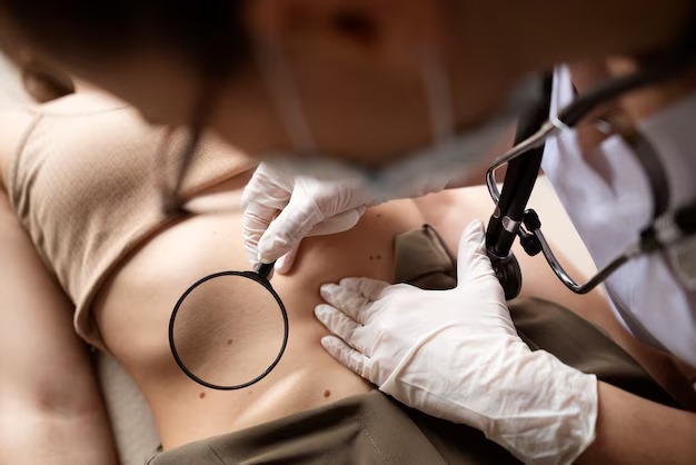
Moles, also known as nevi, are common skin growths that most people have. While the majority of moles are harmless, some can develop into melanoma, a serious form of skin cancer. The challenge for healthcare providers is to accurately distinguish between benign moles and potentially malignant ones. Two primary methods are used for mole evaluation: visual inspection and dermoscopy. This article explores the differences between these methods, the importance of Dermoscopy Mole Evaluation in Dubai, and why dermoscopy is increasingly favored for early detection of skin cancer.
Understanding Mole Evaluation
What Are Moles?
Moles are clusters of pigmented cells that appear as small, dark spots on the skin. They can vary in color, shape, and size, and are usually benign. However, changes in a mole’s appearance can sometimes indicate the development of melanoma, making regular monitoring essential.
Why Evaluate Moles?
Evaluating moles is crucial for early detection of melanoma. The earlier melanoma is identified, the better the chances of successful treatment. Regular skin checks and professional evaluations are key to catching potential problems early.
Methods of Mole Evaluation
There are two main methods for evaluating moles: visual inspection and dermoscopy. Each method has its advantages and limitations, and understanding these can help in choosing the most appropriate approach for mole assessment.
Visual Inspection: The Traditional Approach
What Is Visual Inspection?
Visual inspection is the traditional method used by healthcare providers to assess moles. It involves examining the mole with the naked eye, looking for signs that could indicate malignancy, such as asymmetry, irregular borders, multiple colors, large diameter, and any changes over time.
The ABCDEs of Mole Evaluation
The ABCDE rule is a common guideline used during visual inspections:
- A for Asymmetry: One half of the mole does not match the other.
- B for Border: The edges are irregular, ragged, or blurred.
- C for Color: The color is not uniform and may include shades of brown, black, or even red.
- D for Diameter: The mole is larger than 6mm (about the size of a pencil eraser).
- E for Evolving: The mole changes in size, shape, or color over time.
These criteria help in identifying moles that may require further investigation or removal.
Limitations of Visual Inspection
While visual inspection is a valuable tool, it has its limitations. Subtle changes in a mole or early signs of melanoma can be difficult to detect with the naked eye. Additionally, benign moles can sometimes resemble malignant ones, leading to unnecessary biopsies or, conversely, missed diagnoses. This is where dermoscopy plays a critical role in improving accuracy.
Dermoscopy: A Closer Look
What Is Dermoscopy?
Dermoscopy, also known as dermatoscopy, is a non-invasive diagnostic technique that involves using a handheld device called a dermatoscope. This device magnifies the skin and uses polarized light to allow for a more detailed examination of moles. Dermoscopy provides a view of subsurface skin structures that are not visible to the naked eye.
How Dermoscopy Enhances Evaluation
Dermoscopy enables the visualization of patterns and structures within the mole, such as pigment networks, vascular structures, and the arrangement of colors. These features can provide critical clues about whether a mole is benign or malignant. The increased detail allows for a more accurate assessment, reducing the likelihood of false positives or negatives.
Dermoscopy in Practice
In clinical practice, dermoscopy has become an essential tool for dermatologists and other healthcare providers. It is particularly useful for evaluating atypical moles and identifying early signs of melanoma that might be missed during a visual inspection. Studies have shown that dermoscopy can significantly improve diagnostic accuracy, leading to earlier and more effective treatment of skin cancer.
Comparing Dermoscopy and Visual Inspection
Accuracy and Reliability
Dermoscopy offers a higher level of accuracy compared to visual inspection alone. By providing a detailed view of a mole’s structure, dermoscopy reduces the risk of misdiagnosis. This increased accuracy is especially important in detecting early melanomas, which can be challenging to identify based solely on visual inspection.
Patient Safety and Confidence
With dermoscopy, patients benefit from a more thorough and reliable evaluation of their moles. This can lead to fewer unnecessary biopsies and a greater sense of security in knowing that their skin is being monitored with the best available technology. For healthcare providers, dermoscopy offers a more precise tool for assessing moles, ultimately improving patient outcomes.
When Is Visual Inspection Still Useful?
While dermoscopy is a powerful tool, visual inspection remains a valuable first step in mole evaluation. It is quick, non-invasive, and can be easily performed during routine skin checks. For many patients, a visual inspection may be sufficient to determine whether further examination is needed. However, for moles that are atypical or have changed over time, dermoscopy is often the next logical step.
The Importance of Accurate Mole Evaluation
Early Detection Saves Lives
The primary goal of mole evaluation is the early detection of melanoma. Melanoma is the deadliest form of skin cancer, but it is highly treatable when caught early. Accurate mole evaluation, whether through visual inspection or dermoscopy, plays a critical role in identifying melanoma before it spreads.
Reducing Unnecessary Procedures
Accurate evaluation also helps reduce unnecessary procedures. By distinguishing between benign and malignant moles more effectively, dermoscopy can prevent unnecessary biopsies and surgeries, which can be stressful and costly for patients.
Empowering Patients Through Education
Educating patients about the importance of regular skin checks and the differences between visual inspection and dermoscopy empowers them to take an active role in their skin health. Understanding the tools available for mole evaluation can help patients make informed decisions about their care and seek out the best possible screening methods.
Conclusion: Choosing the Right Method for Mole Evaluation
When it comes to mole evaluation, both visual inspection and dermoscopy have their place. Visual inspection is a quick and accessible method that can identify moles needing further examination, while dermoscopy offers a more detailed and accurate assessment, particularly for atypical or suspicious moles. The importance of accurate mole evaluation cannot be overstated, as early detection of melanoma is crucial for successful treatment. For patients and healthcare providers alike, understanding the strengths and limitations of each method can lead to better decision-making and ultimately, better outcomes in the fight against skin cancer.


Comments