What is POCT?
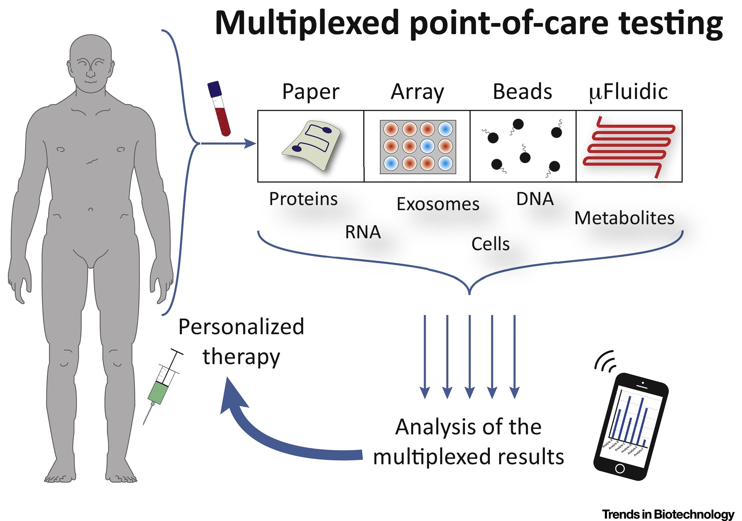
 Multiplexed point-of-care testing
POCT (Point-of-Care Testing) refers to clinical and bedside testing conducted in close proximity to patients. It is often not performed by clinical laboratory personnel. POCT holds significant importance in disease prevention, etiology determination, prognosis, enhancing treatment outcomes, and reducing healthcare expenses. It meets clinical testing needs across various healthcare settings. POCT serves as a complement to traditional laboratory testing methods. It is characterized by short testing times, spatial flexibility, and simple operation.
However, as it is decentralized, the accuracy of test results in POCT can be greatly affected by non-standardized procedures and inadequate quality control. The field of POCT products has advanced through four generations: qualitative, semi-quantitative, fully quantitative, and automated, due to the continuous innovation in testing technologies. POCT technical principles include dry chemistry, colloidal gold, immunofluorescence, microfluidic chips, chemiluminescence, biosensors, and biochips.
Colloidal gold is the most commonly used nanoparticle for optical labeling due to its ease of use, biocompatibility, and high visibility. It appears deep red in color, which can cause confusion in on-site testing as it displays the same color in two or more test lines. Polystyrene particles (PS particles), also referred to as latex beads (LBs), impregnated with various bright dyes have several advantages. They are easy to synthesize and have multi-color capability, making them an ideal choice for developing robust immunochromatographic analysis.
CD Bioparticles offers a variety of uniform and monodisperse colored polystyrene particles, including Diagpoly™ Colored Polystyrene Particles. These particles are highly pigmented, which can improve visual detection in assays such as latex agglutination, dipstick, and membrane-based assays.
Advantages of Colored Polystyrene Particles for POCT
- Simplicity and speed: In contrast to colloidal gold and fluorescent nanoparticles, colored polystyrene nanoparticles can be prepared by copolymerization with dyes, solvent swelling of templated nanoparticles and self-assembly of block copolymers and dyes, providing various surface functional groups. In addition, polystyrene nanoparticles exhibit excellent biocompatibility and flexibility, facilitating effective conjugation with specific proteins and antibodies.
- Increased sensitivity and specificity: Each particle of colored polystyrene nanoparticles is carefully designed in size and coated with specific ‘capture molecules’ designed to trap their intended targets. When samples containing target molecules are introduced, these coated particles act like magnets, attracting their specific targets with high affinity. This targeted binding significantly reduces the chance of capturing the wrong molecules, thus minimizing false positives. The large surface area of the polystyrene particles allows more capture molecules to be accommodated, increasing test sensitivity and minimizing false negatives. Polystyrene itself plays a critical role in detection accuracy. This robust material is chemically inert, meaning it does not interfere with the binding process or produce false signals. In addition, its uniform surface provides a stable platform for capture molecule attachment, ensuring consistent and reliable results.
- Versatility and multiplex detection: While LFTs are typically designed for the rapid detection of single analytes per assay, there is growing interest in multiplex analysis due to its potential for increased throughput, reduced sample requirements and improved assay efficiency. The most common approach to achieving multiplex detection in LFTs is to use multiple test lines on the test strip, each reporting the detection results of specific analytes. Although colloidal gold has a distinct red color, it is limited to single color testing and is not suitable for multiplexing. In contrast, colored latex beads offer a variety of vivid colors for easier visual assessment of multiple test lines, reducing the risk of misinterpretation of results.
- Cost-effectiveness: Test strips produced with colored latex beads from different batches have relatively little variation between batches, and production processes typically require minimal or no adjustment. Colloidal gold readily adsorbs to certain materials, even when glass containers are used, which introduces unavoidable risks in large-scale production. Therefore, a more cautious approach is to opt for colored latex particles. The use of colored latex particles in the production of immunochromatographic test strips is therefore more cost effective.
- Customization and flexibility: Through unique surface modification processes, CD Bioparticles can add different functional groups to the surface of colored latex particles, such as carboxyl and amine groups, to facilitate antibody/antigen coupling on microsphere surfaces. Carboxyl-modified colored latex particles introduce more N and O atoms onto the microsphere surface, increasing the rate of antibody migration to the material surface and effectively improving the coupling efficiency. Our microspheres feature uniform particle sizes, stable and controllable surface functional groups and excellent experimental reproducibility. CD Bioparticles can customize colored latex particles with various functional group surface modifications according to customer requirements.
Features of CD Bioparticles’ Colored PS Particles
- Our monodisperse-colored polystyrene latex microspheres have perfect sphericity and narrow size distribution. With a wide size range from 30 nm to 750 µm, they have larger diameters compared to colloidal gold particles typically used in immunochromatography products (colored latex microspheres from 200 nm to 400 nm, colloidal gold particle sizes from 20-40 nm), offering higher relative sensitivity.
Shop for DiagPoly™ Colored Polystyrene Particles, 200 nm to 400 nm.
- We offer a variety of vividly colored latex particles including black, blue, green, orange, red, purple and yellow. With enhanced coloring capabilities, results are more visually intuitive, allowing for easy visual interpretation or multiplexed analysis.
Black Latex ParticlesRed Latex ParticlesBlue Latex ParticlesViolet Latex ParticlesGreen Latex ParticlesYellow Latex ParticlesOrange Latex Particles
- These colored microspheres are manufactured using an internal embedding method, where the dye is located inside the microsphere, ensuring minimal leakage and stable structural integrity. Our products exhibit exceptional stability in physicochemical properties, which facilitates easy storage.
- With a high specific surface area and negative surface charge, our colored latex particles can be covalently linked to antibodies via chemical reactions or adsorbed for antibody conjugation. For molecules containing multiple sulphur atoms, latex microspheres offer superior labelling effects. The polystyrene matrix exhibits excellent stability and affinity, as well as good biocompatibility.
- CD Bioparticles has achieved scale production, offering product specifications from 10 mL to 1000 mL with consistent performance, uniform particle size and minimal variation between batches.
Applications of Colored PS Particles
- Temperature sensing is a promising approach to improve the detection sensitivity of lateral flow immunoassays (LFIA) for POCT. By optically stimulating colored latex beads (CLB) on LFIA strips with laser power below 100 mW, temperature elevations above 100 °C can be easily achieved. The researchers observed a dependence of membrane-bound CLB density on temperature elevation and found that the light absorption of red (and black) latex beads at 520 nm increased 1.3-fold (and 3.2-fold, respectively). This increase is attributed to multiple light scattering in this highly porous medium, a mechanism that could significantly impact the sensitivity improvement of LFIA. The detection limit is 1 × 10^5 particles/mm².
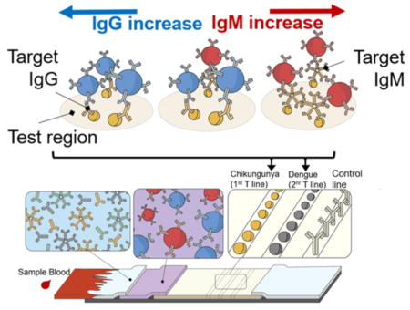
- The researchers proposed a novel lateral flow detection scheme based on two-color latex labels (blue latex beads, 400 nm, and red latex beads, 400 nm) for multiplex detection of CHIKV and DENV IgG/IgM on a single lateral flow detection strip. They demonstrate that their detection can indicate the presence of anti-CHIKV IgG/IgM and anti-DENV IgG/IgM by observing the color and position of the test areas for CHIKV and DENV.

Two-color lateral flow assay for multiplex IgG/IgM detection of several febrile illnesses
According to the World Health Organization’s (WHO) REASURED guidelines, an ideal point-of-care testing should have real-time connectivity, ease of specimen collection, affordable, sensitive and specific, user friendly, rapid and robust, equipment-free and environment friendly, and deliverable to end-users. Lateral flow testing will therefore remain an important format for future point-of-care devices. In particular, multiplexing capabilities could represent the next generation of POC testing. One way to achieve multiplex detection in LFTs is to use uniquely labeled or differently colored detector particles (nanoscale beads responsible for visualizing signals on test strips). CD Bioparticles’ colored polystyrene beads significantly increase detection sensitivity and reduce cost, facilitating multiplexing in lateral flow applications.
References
Lee, S., Mehta, S., & Erickson, D. (2016). Two-color lateral flow assay for multiplex detection of causative agents behind acute febrile illnesses. Analytical chemistry, 88(17), 8359-8363.
Azuma, T., Hui, Y. Y., Chen, O. Y., Wang, Y. L., & Chang, H. C. (2022). Thermometric lateral flow immunoassay with colored latex beads as reporters for COVID-19 testing. Scientific Reports, 12(1), 3905.


Comments