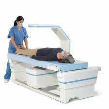
In the realm of modern medicine, diagnostic technologies play a pivotal role in identifying health conditions and guiding appropriate treatments. Among these, Dual-Energy X-ray Absorptiometry (DEXA) scan stands out as a crucial tool in assessing bone health. Utilized primarily to diagnose osteoporosis, DEXA scanning offers valuable insights into bone density, aiding in early intervention and preventive measures. This article aims to delve into the significance, procedure, and implications of DEXA scanning in healthcare.
What is DEXA Scan?
DEXA scan, also known as bone densitometry, employs X-ray technology to measure bone mineral density (BMD) and determine bone strength. It is considered the gold standard for assessing skeletal health due to its precision, minimal radiation exposure, and non-invasiveness. The procedure involves passing low-dose X-ray beams through the bones, measuring the amount of energy absorbed. Based on this data, healthcare providers can evaluate bone density and assess the risk of fractures.
Significance of DEXA Scan
Osteoporosis, characterized by weakened bones prone to fractures, poses a significant health concern, particularly among the elderly and postmenopausal women. DEXA scan plays a crucial role in diagnosing osteoporosis by detecting low bone density before fractures occur. Early detection enables timely intervention through lifestyle modifications, dietary supplements, or medication to prevent debilitating fractures and improve quality of life.
Procedure:
The DEXA scanning procedure is quick, painless, and typically performed on an outpatient basis. Patients lie flat on a table while a mechanical arm passes over the body, emitting X-rays focused on specific skeletal sites, such as the spine, hip, or forearm. Measurements are taken at these sites, and the results are compared with reference values to assess bone density. The entire process usually takes around 10 to 30 minutes, depending on the areas being scanned.
Interpreting Results:
DEXA scan results are presented as T-scores and Z-scores, indicating deviations from the average bone density of a healthy young adult. T-scores compare bone density with that of a healthy young adult of the same gender, highlighting the risk of osteoporosis:
- Normal: T-score above -1
- Osteopenia (Low bone density): T-score between -1 and -2.5
- Osteoporosis: T-score of -2.5 or lower
Z-scores, on the other hand, compare bone density with individuals of the same age, gender, and size. These scores help assess bone health in younger individuals and children.
Clinical Applications:
Beyond diagnosing osteoporosis, DEXA scanning finds application in various clinical scenarios:
- Monitoring Treatment: DEXA scans track changes in bone density over time, allowing healthcare providers to assess the effectiveness of osteoporosis treatments and modify management strategies accordingly.
- Risk Assessment: Individuals with certain risk factors, such as family history of osteoporosis, prolonged steroid use, or chronic conditions impacting bone health, may benefit from periodic DEXA scans to assess fracture risk.
- Research: DEXA scanning serves as a valuable tool in research studies investigating the efficacy of new therapies, understanding bone metabolism, and elucidating factors influencing bone density.
Conclusion:
In the landscape of preventive medicine, DEXA scan emerges as a cornerstone in the assessment of bone health, offering valuable insights into bone density and fracture risk. Its role extends beyond diagnosis, encompassing monitoring, risk assessment, and research endeavors aimed at enhancing our understanding of skeletal health. As we strive towards proactive healthcare, the significance of DEXA scanning in promoting musculoskeletal well-being cannot be overstated, emphasizing the importance of integrating this technology into routine clinical practice.


Comments