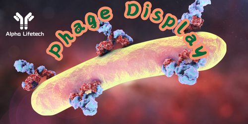
Phage display is a technique used to study protein interactions and to identify peptides, proteins, or antibodies that have a high affinity for specific target molecules. This technique leverages the ability to fuse foreign peptides or proteins with phage-coat proteins, allowing the foreign proteins to be displayed on the phage surface. Below is a standard protocol for performing a phage display experiment.
Materials Needed:
1. Phage Display Vector: A vector that contains the phage coat protein gene (e.g., gene III for M13 phage).
2. Insert DNA: The DNA encoding the peptides or proteins to be displayed.
3. Host Bacteria: Escherichia coli strains such as XL1-Blue, TG1, or ER2738.
4. Helper Phage: M13KO7 or similar, used to produce phagemid particles.
5. LB Agar Plates: Containing an antibiotic for selection (e.g., ampicillin).
6. Growth Medium: SOC medium, LB medium with antibiotics.
7. Reagents: PEG/NaCl for phage precipitation, PCR reagents, restriction enzymes, ligase, and buffers.
8. Equipment: Thermocycler, incubator, spectrophotometer, centrifuge, shaker.
Protocol Steps:
1. Preparation of DNA Insert and Vector:
Amplification of Insert DNA: Use PCR to amplify the DNA encoding the peptide or protein of interest. Primers should include restriction sites for cloning into the phage display vector.
Purification of PCR Product: Use a gel extraction kit to purify the PCR product.
Digestion of Vector and Insert: Digest both the phage display vector and insert DNA with the appropriate restriction enzymes to create compatible ends for ligation.
Purification: Purify the digested vector and insert using a DNA purification kit.
2. Ligation and Transformation:
Ligation: Set up a ligation reaction with the digested vector and insert using T4 DNA ligase. Incubate the reaction mixture at 16°C overnight or at room temperature for a few hours.
Transformation: Transform the ligated DNA into competent E. coli cells using a heat shock method or electroporation.
Recovery: Incubate the transformed cells in SOC medium at 37°C with shaking for 1 hour.
Plating: Plate the transformed cells on LB agar plates containing the appropriate antibiotic (e.g., ampicillin) to select colonies containing the phage display vector. Incubate the plates overnight at 37°C.
3. Phage Rescue:
Infection with Helper Phage: Pick a single colony and inoculate it into an LB medium with an antibiotic. Grow until OD600 reaches 0.5, then infect the culture with helper phage (e.g., M13KO7) at a multiplicity of infection (MOI) of 20:1.
Incubation: Incubate the infected culture at 37°C for 30 minutes without shaking, followed by shaking incubation at 250 rpm for 1 hour.
Amplification: Add antibiotics (e.g., kanamycin) to select for cells infected with helper phage. Incubate at 30°C overnight with shaking.
4. Phage Purification:
Collection: Centrifuge the overnight culture at 10,000 x g for 10 minutes to pellet the cells. Collect the supernatant containing the phage particles.
Precipitation: Add PEG/NaCl to the supernatant to precipitate the phage. Incubate on ice for 1 hour.
Centrifugation: Centrifuge the mixture at 10,000 x g for 15 minutes. Discard the supernatant and resuspend the phage pellet in a suitable buffer (e.g., PBS).
5. Phage Titering:
Titering: Perform serial dilutions of the purified phage and infect a fresh culture of E. coli. Plate the infected cells on LB agar plates with antibiotics to determine the titer of the phage library.
6. Biopanning (Selection for High-Affinity Binders):
Coating the Target: Coat a microtiter plate or magnetic beads with the target protein or antigen.
Incubation with Phage Library: Add the phage library to the coated target and incubate to allow binding. Non-specific binding sites can be blocked using BSA or casein before adding the phage.
Washing: Wash the wells or beads several times with washing buffer (e.g., PBS with 0.1% Tween-20) to remove unbound and weakly bound phage.
Elution: Elute the bound phage using an elution buffer (e.g., low pH buffer, trypsin) to disrupt the interaction between the phage and the target.
Amplification: Infect fresh E. coli cells with the eluted phage and amplify the enriched phage population as described in the phage rescue step.
7. Additional Rounds of Biopanning:
Repeat the biopanning process for 2-4 rounds to further enrich for high-affinity binders. Each round will increasingly select for phage that binds with high specificity and affinity to the target.
8. Screening and Characterization:
Screening: After the final round of biopanning, pick individual phage clones and screen for binding activity to the target using ELISA or other suitable assays.
Sequencing: Sequence the DNA of positive clones to identify the peptide or protein sequences responsible for binding.
9. Validation:
Confirm the binding affinity and specificity of the selected phage-displayed peptides or proteins using additional biochemical and biophysical assays such as surface plasmon resonance (SPR), isothermal titration calorimetry (ITC), or competitive binding assays.
Conclusion:
Phage display is a robust method for identifying and characterizing peptides, proteins, or antibodies with high affinity for specific targets. By following the outlined protocol, researchers can construct a phage display library, perform selection, and identify strong binders for further development in research, diagnostics, or therapeutic applications.


Comments